


English
English

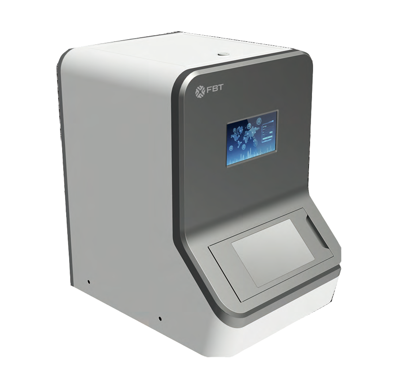
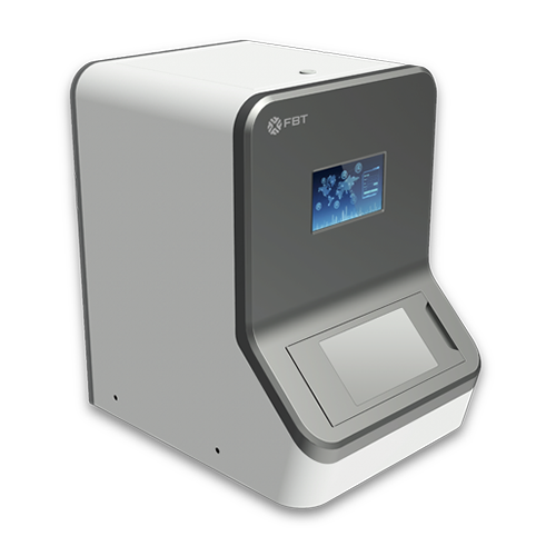
The Clippers series uses professional linear cameras with independent color processing channels to ensure more natural and accurate color reproduction and image clarity, and can easily obtain high-precision, high-quality panoramic digital slice images. One-key operation, simple and convenient.
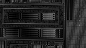
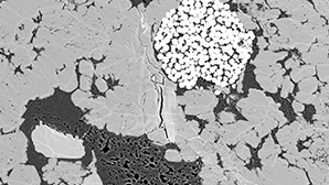
Photoelectric combination: The high-definition map scanner provide precise location to correlate and overlap the SEM coordinates. And its optical image can be used as a NavigatorSEM to seamlessly connect with SEM image. Therefore, one can quickly and accurately locate and observe designated locations through the SEM.
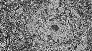
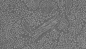
We apply mature industrial scanner technology in the semiconductor industry to life sciences, materials science, failure analysis, research and other fields. Mainly used in the research and production of ultra-large biological mesoscopic tissue structure imaging and tissue depth information, high-throughput cross-scale material characterization, large-scale sample defect screening, coating process detection, flat panel display detection, automatic particle screening, and process control.
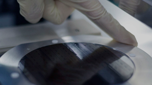
The friendly human-computer interface makes it easy for users to input and take out samples, and real-time system status monitoring can be realized through it.


Sample damage is reduced with high-sensitivity camera, precisely calibrated optical system and soft illumination. Independent color processing channel ensures more natural and accurate color reproduction and image clarity
| Parameter type | Value |
| Resolution | ≤7um/pixel |
| Imaging distortion | 0.01% |
| Wavelength range | 350nm-1100um |
| Working distance | 50mm±5mm |
| Sample table | Sample movement range:Y≥210(mm ) Z≥20(mm) Repetitive positioning accuracy:≤±500nm Straightness:±10um/300mm |
| camera | High-throughput, low-noise camera Maximum Pixels:16K Sensing mode: CMOS Connection mode: Standard Cameralink interface Pixel size:3.5 μm x 3.5 μm Dimension:76.0 mm x 76.0 mm x 30.7 mm Work Temperature:0 °C to 65 ° |
| Digital image processing | Total imaging time:9s Image resolution:16kX 32k Positioning error:8um |
| Clipper software | Optical linescanner automatic image acquisition software Coordinate transformation software for nanoscope and electron microscope Image measurement software |
| Computer hardware configuration (optional) | CPU: Intel core i5-8500 memory:8G Hard drive capacity:4T Graphics card: Graphics card Maximum video memory setting4G Optical drive: writerable optical drive Display:24 inch wide screen LED backlight |


 View video
 Product Introduction
 Feature and Benefits
 Technical Specifications
 Related applications and cases